Eye Examinations: Understanding the Process
Eye investigations are critical for preserving and improving vision. Regular check-ups can help detect issues early, allowing for timely treatment and prevention of more serious conditions. If you experience any changes in your vision, it is important to consult an eye care professional as soon as possible.
Now, I’ll create three individual photos to complement this article:
- A visual acuity test with the Snellen chart.
- A slit lamp exam in progress.
- A close-up of an ophthalmoscope during an eye examination.
Eye Investigations: Understanding the Process
The human eye is a remarkable organ, responsible for one of the most important senses: vision. Eye investigations are essential in diagnosing and treating various eye conditions, maintaining good eye health, and preventing vision loss. These investigations range from simple eye exams to more complex diagnostic tests using advanced technologies. Let’s explore some of the common eye investigations and their importance.
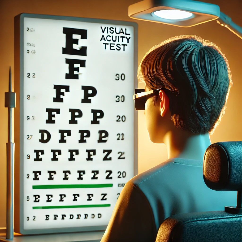
1. Visual Acuity Test
A visual acuity test is the most common eye examination, often performed during routine eye check-ups. This test measures how well you can see the details of a letter or symbol from a specific distance. The standard chart used is the Snellen chart, featuring rows of letters that progressively get smaller.
Why It’s Important:
- Detects issues like nearsightedness (myopia), farsightedness (hyperopia), and astigmatism.
- Helps determine whether corrective lenses or further treatment are needed.
2. Slit Lamp Examination
A slit lamp is a powerful microscope with a bright light that enables the ophthalmologist to examine the front part of the eye, including the cornea, iris, lens, and anterior chamber. It provides a magnified view, allowing for a detailed inspection of these areas.
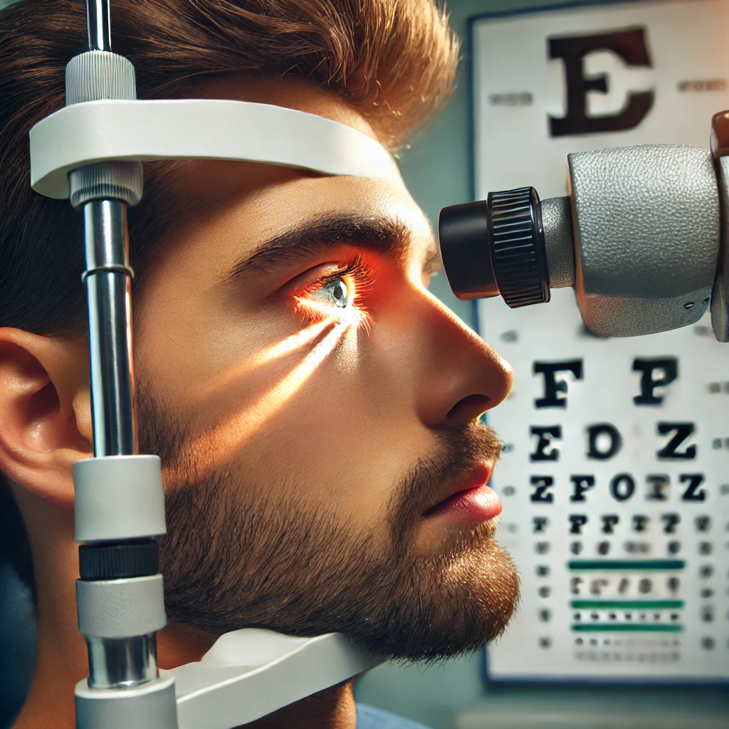
Why It’s Important:
- Used to detect issues such as cataracts, corneal injuries, and infections.
- Helps in identifying early signs of eye diseases like glaucoma and macular degeneration.
3. Ophthalmoscopy
Ophthalmoscopy (also called fundoscopy) is an exam that allows a doctor to see the back of the eye, including the retina, optic disc, and blood vessels. This is typically done using an ophthalmoscope, a hand-held instrument that shines a light into the eye.
Why It’s Important:
- Helps in diagnosing conditions like diabetic retinopathy, retinal detachment, and optic nerve damage.
- Provides critical insights into overall eye health and possible systemic diseases like hypertension and diabetes.
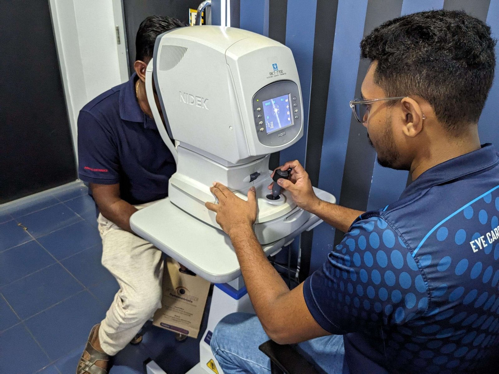
4. Tonometry
Tonometry is used to measure the pressure inside the eye (intraocular pressure or IOP), which is a critical test for diagnosing glaucoma, a disease that can cause irreversible blindness. There are various types of tonometers, including the non-contact (air puff) and Goldmann applanation tonometry.
Why It’s Important:
- High intraocular pressure can be an early indicator of glaucoma.
- Regular monitoring is crucial for patients at risk of developing this condition.
5. Optical Coherence Tomography (OCT)
OCT is an advanced imaging technique that provides detailed cross-sectional images of the retina. It is often used to diagnose and monitor diseases of the retina and optic nerve. This non-invasive test allows the doctor to see each of the retina’s layers and measure their thickness.
Why It’s Important:
- Essential for diagnosing macular degeneration, diabetic retinopathy, and glaucoma.
- Offers precise monitoring of retinal health over time.