DIAGNOSTIC TESTS

Diagnostic Tests: Essential Tools for Eye Health
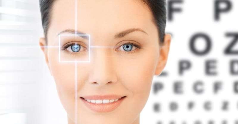
Eye diagnostic tests are crucial in detecting and monitoring various eye conditions that can affect vision and overall eye health. These tests help eye care professionals diagnose diseases early, allowing for timely treatment and prevention of further complications. Below, we explore five key diagnostic tests, each addressing different aspects of eye health.

1. Dry Eye Assessment
Dry eye syndrome occurs when your eyes do not produce enough tears or when the tears evaporate too quickly. A dry eye assessment helps determine the severity of the condition and its underlying causes.
Common Dry Eye Tests:
- Tear Break-Up Time (TBUT): Measures how quickly the tear film breaks up on the surface of the eye. A faster break-up time indicates dry eye.
- Schirmer’s Test: A small strip of paper is placed under the lower eyelid to measure tear production. The amount of moisture on the strip helps assess tear volume.
- Ocular Surface Staining: Dyes like fluorescein are applied to the eye to observe dry spots on the cornea, indicating damage from insufficient tear coverage.
Why It’s Important:
- Early detection helps prevent damage to the cornea and discomfort from chronic dry eye.
- Tailored treatment can involve artificial tears, lifestyle changes, or medications to improve tear production and retention.
2. Glaucoma Assessment
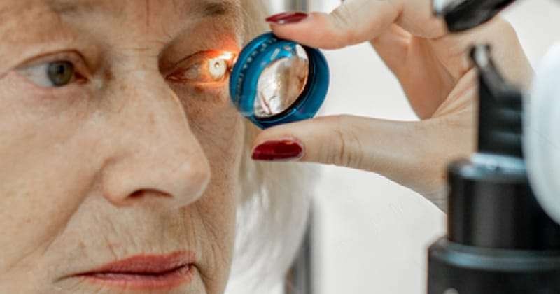
Glaucoma is a group of diseases that damage the optic nerve, often caused by elevated intraocular pressure (IOP). It is a leading cause of blindness if not treated early. A comprehensive glaucoma assessment involves several diagnostic tests to evaluate the condition.
Common Glaucoma Tests:
- Tonometry: Measures intraocular pressure (IOP) to detect ocular hypertension, a major risk factor for glaucoma.
- Visual Field Test: Assesses peripheral vision loss, one of the early signs of glaucoma.
- Optic Nerve Evaluation: A dilated eye exam allows the doctor to assess the optic nerve for signs of damage.
Why It’s Important:
- Glaucoma often progresses without symptoms until significant vision loss has occurred. Regular screenings and early intervention are key to preventing irreversible damage.
3. Diabetic Retinopathy Assessment
Diabetic retinopathy is a complication of diabetes that affects the blood vessels of the retina, leading to vision impairment or loss. A diabetic retinopathy assessment is essential for individuals with diabetes to monitor and manage this condition.
Common Tests for Diabetic Retinopathy:
- Dilated Fundus Exam: After dilating the pupils, the doctor examines the retina for abnormalities like leaking blood vessels, swelling, or new blood vessel growth.
- Fluorescein Angiography: A dye is injected into the bloodstream, and photographs are taken to show blood flow and detect any leakage in the retinal vessels.
Why It’s Important:
- Early detection of diabetic retinopathy allows for timely treatment, such as laser therapy or injections, to slow the progression and protect vision.
4. Optical Coherence Tomography (OCT)

OCT is a non-invasive imaging test that provides detailed cross-sectional images of the retina. It is often used to assess the macula, optic nerve, and retina for various eye conditions.
How OCT Works:
- The OCT scan uses light waves to capture 3D images of the retina’s layers, allowing for precise measurement of their thickness. This can reveal swelling, thinning, or fluid buildup, which are indicative of diseases like age-related macular degeneration (AMD), glaucoma, or diabetic retinopathy.
Why It’s Important:
- OCT provides an in-depth look at the retina, offering early detection and monitoring of retinal and optic nerve conditions.
- It is essential for tracking the progression of chronic eye diseases and evaluating treatment effectiveness.
5. Fundus Photography
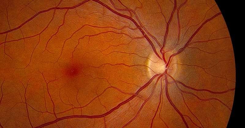
Fundus photography involves taking detailed images of the back of the eye (the fundus), which includes the retina, optic disc, and macula. These high-resolution photographs allow for a thorough assessment of the eye’s internal structures.
What Fundus Photography Detects:
- Retinal detachment.
- Macular degeneration.
- Diabetic retinopathy.
- Glaucoma damage.
Why It’s Important:
- The images serve as a record for tracking changes in the eye over time, which is especially useful for chronic conditions like diabetic retinopathy or glaucoma.
- Early diagnosis of diseases through fundus photography can lead to better treatment outcomes.